
(© appledesign - stock.adobe.com)
FREIBURG, Germany — Heart disease could be detected years in advance in high-risk patients thanks to next-generation scanning technology. Called ultra-high-resolution computed tomography (UHR-CT), this new type of imaging provides excellent quality and precise diagnosis of coronary artery disease non-invasively, identifying threats years before they manifest.
Technology used today — coronary computed tomography angiography (CCTA) — is quite effective at ruling out coronary artery disease in patients with low or intermediate risk. However, screening high-risk individuals can be challenging due to a high prevalence of calcifications, which can often appear more extensive than they truly are and result in false positives.
“Consequently, patients may undergo unnecessary, often invasive, testing. This is the reason why current guidelines do not recommend using CCTA in high-risk individuals,” says lead author Dr. Muhammad Hagar from the University of Freiburg, Germany, in a media release.
This innovative tool employs a photon-counting detector. In the first comprehensive study of its kind, researchers compared its diagnostic accuracy with the reference standard of invasive coronary angiography (ICA) in 68 patients suffering from severe aortic valve stenosis, a common but serious valve disease that impedes or blocks blood flow from the heart.
The UHR-CCTA proved to be highly sensitive and specific for coronary artery disease detection, achieving a median overall image quality score of 1.5 on the 5-point Likert scale (where one represents excellent and five signifies non-diagnostic). Approximately 80% of segments were rated as good or excellent, indicating that this technique could soon be available to high-risk patients.
“It appears that the spectrum of patients benefiting from undergoing non-invasive CCTA has been significantly broadened by photon-counting detector technology. This is excellent news for these patients and the imaging community,” says Dr. Hagar.
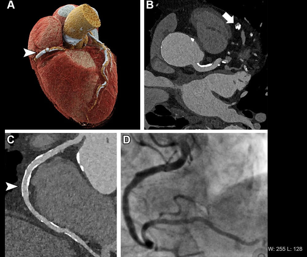
(credit: Radiological Society of North America)
The high resolution of UHR-CCTA results from an increased number of emitted photons, but this also leads to greater radiation exposure compared to conventional CT scanners. Currently, the technology is in its early stages, and researchers are developing methods to decrease radiation exposure.
“Currently, the technique is feasible for high-risk patients, in whom the benefits outweigh the risks, but should not be applied to all patients referred for cardiac CT imaging,” adds Dr. Hagar.
Photon-counting CT is relatively rare worldwide, but experts predict the technology will become more prevalent within the next decade.
“At the University of Freiburg, we had the privilege to work with the technology since its introduction and I am convinced that photon-counting CT is the beginning of a new generation of CT scanners, similar to the introduction to multislice CT 30 years ago,” says Dr. Hagar.
The team is now investigating the diagnostic potential of photon-counting CT technology in other clinical scenarios such as oncological imaging, and expanding their research to include groups for whom CT imaging is currently not feasible, like patients with coronary stents. They are also exploring the potential for heart muscle assessment with photon-counting CT. Early results suggest the technology could improve soft tissue resolution, greatly benefiting disease characterization.
“For me, these are exciting times, and it is just great to be part of a very active group working with this new technology,” concludes Dr. Hagar.
This research is published in the journal Radiology.
South West News Service writer Mark Waghorn contributed to this report.


At what age should you be checked for your heart?